Opening a can of worms
10 October 2014 - Biochemical
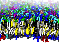 The image of the week is the result of a molecular dynamics simulation of a mammalian plasma membrane (DOI). It looks like a can of worms has opened but the piece of cell wall depicted (a bilayer) consists of a complex mixture of 63 different worm-like lipids (phosphatidylcholines, sphingomyelin and gangliosides for the inside and hosphatidylethanolamine and phosphatidylserine for the outside) with additional cholesterol and water thrown in. This process is called computational microscopy.
The details: simulations were performed using MARTINI and using a tool called INSANE (INSert membrANE), around half a million particles (lipids, water molecules and counter ions) were crammed into a box measuring 70 by 70 by 11 nm and monitored for 40 microseconds. Initially all players are distributed randomly but phase-separation takes place instantly with cholesterol congregating and also aliphatic lipid tails in general. The gangliosides were also found to form clusters.
The simulation reveals how cholesterol-rich regions flip-flop between the in- and outside. In the accompanying video these regions are like waves in an ocean. Not able to find: the amount of time the computations took, hours?, days?, weeks?. The 40 microseconds were not enough for everything to equilibrate. Hence the curiosity.